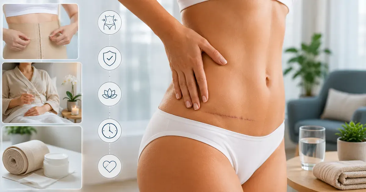
What Is Fibrosis After Liposuction?
Fibrosis refers to the formation of excess fibrous connective tissue — essentially scar tissue — in response to injury. After liposuction, the mechanical disruption of fat cells, cannula movement through tissue, and removal of fat creates a controlled injury that the body heals through its normal inflammatory cascade.
Most patients notice fibrosis as:
- Firm or hard patches beneath the skin in treated areas
- Lumpy or uneven texture that can be felt but may not always be seen
- Tightness or pulling sensation when moving
- Areas that feel stiffer than expected given the passage of time
This is distinct from the normal swelling of the first few days, which is fluid-based. Fibrosis is a tissue-level process — the scar matrix forms in the space where fat was removed.
Why Liposuction Specifically Causes Fibrosis
Liposuction is uniquely fibrosis-prone compared with other cosmetic procedures because of how the cannula moves through tissue. During fat removal, the surgeon makes repeated back-and-forth passes through the subcutaneous fat layer. Each pass disrupts not only fat cells but also the fine connective tissue septa — the fibrous bands that run between fat lobules — and causes micro-tears in small blood vessels and lymphatic channels. The cumulative mechanical trauma across dozens or hundreds of cannula passes creates a wide zone of injury rather than a single incision line. The body cannot simply "stitch" this zone closed the way it does a surgical cut; it must fill the entire treated volume with a provisional repair matrix.
That provisional matrix is collagen — secreted by fibroblasts that migrate into the disrupted area in response to chemical signals released by damaged cells and platelets. In the early weeks, this collagen is deposited rapidly and in a disorganised pattern: dense, cross-linked, and stiff. It is this dense, immature collagen network that patients feel as the firmness and lumps of fibrosis. Over subsequent months, specialised cells called myofibroblasts and matrix metalloproteinases (MMPs) break down the excess collagen and remodel it into softer, more organised tissue. The extent of fibrosis is therefore directly related to the extent of mechanical trauma — which is why large-volume liposuction, aggressive technique, or multiple treatment areas in a single session tend to produce more pronounced fibrosis than smaller procedures.
When Fibrosis Peaks and When It Resolves
Fibrosis follows a fairly predictable timeline in most patients, though individual variation is significant. The first signs — a diffuse firmness beneath the skin — typically emerge around days 5–14 as early swelling begins to subside and the underlying tissue texture becomes palpable. Fibrosis then intensifies through weeks 2–6, which is the proliferative peak: fibroblast activity is at its highest, new collagen is being deposited faster than it is being broken down, and patients often notice the area feels harder than it did immediately after surgery. This can be alarming if patients are not forewarned — it is normal and does not indicate a problem.
From weeks 6–8 onward, the remodelling phase begins to dominate. Most patients with mild to moderate fibrosis notice meaningful softening between months 2 and 4, with continued gradual improvement through month 6. Patients who developed a seroma, had large-volume liposuction, or experienced a haematoma may find fibrosis persists to months 6–12 — the persistent fluid or blood acts as an ongoing inflammatory stimulus that sustains fibroblast activity. Complete resolution — tissue that feels indistinguishable from the surrounding non-treated area — is realistic for the majority of patients, but it requires time and adherence to compression protocols. Final contour assessment is typically deferred to at least 6 months post-surgery for this reason.
Why Fibrosis Happens After Lipo
Liposuction is, at its core, a controlled wound. The body's response to any tissue injury follows a predictable sequence:
- Inflammation (days 1–5): Blood flow increases to the area. Immune cells arrive to clear cellular debris and dead tissue. This phase produces the heat, swelling, and soreness of early recovery.
- Proliferative phase (days 5–21): Fibroblasts (collagen-producing cells) migrate into the damaged area and begin laying down a collagen matrix. This is what becomes fibrosis — the body's temporary scaffold during repair.
- Remodelling phase (weeks 3 – months 12): The initial dense collagen matrix is gradually broken down and replaced with a more organised, softer tissue. This is when fibrosis softens and resolves in most patients.
Larger liposuction volumes, multiple treatment areas (as in Lipo 360), and any complications (seroma, haematoma) increase the inflammatory signal and therefore the extent of fibrosis.
Normal Healing vs Problematic Fibrosis
It is important to distinguish normal healing fibrosis from complications that require medical attention.
| Normal Fibrosis | Requires Surgeon Contact | |
|---|---|---|
| Texture | Firm, lumpy — feels like dense tissue under skin | Fluid-filled, fluctuant swelling (possible seroma) |
| Temperature | Normal skin temperature | Warm or hot to the touch |
| Pain | Mild tightness; occasional achiness | Increasing tenderness, throbbing, or acute pain |
| Skin appearance | Normal colour | Redness or skin changes around lump |
| Systemic symptoms | None | Fever, nausea, feeling unwell |
| Timeline | Gradually softening over weeks | Growing, not reducing; unchanged at 4+ months |
A large fluid-filled swelling that develops days to weeks after surgery is likely a seroma — a collection of post-surgical fluid — rather than fibrosis. Seromas usually require aspiration (draining) by your surgeon. They are not dangerous but do not resolve on their own.
Prevention: What Actually Helps
No intervention prevents fibrosis entirely — it is a normal healing process. However, several measures can minimise its extent and duration:
Compression Garment
Consistent compression garment wear is the most evidence-supported post-liposuction intervention. Compression reduces the dead space between removed-fat layers and skin, limits fluid accumulation, and applies even pressure that supports organised tissue remodelling rather than disordered scar formation. Published research on compression garments supports this approach.2 Wear the garment as prescribed — do not remove it earlier than your surgeon directs.
Graduated Return to Activity
Excessive early activity increases inflammation and can worsen fibrosis. Following your surgeon's staged return-to-exercise protocol — typically light walking from day 1, no strenuous activity until week 6 — supports orderly healing. A 2017 study on activity restriction after liposuction confirms this guidance.1
Lymphatic Massage (MLD)
Manual lymphatic drainage is widely recommended after liposuction. Research on MLD in post-liposuction patients suggests it reduces early fluid accumulation and improves comfort.3 Because persistent interstitial fluid is a key driver of ongoing fibroblast activation, clearing that fluid promptly with MLD may limit the ultimate extent of fibrosis — though direct evidence for MLD reversing established fibrosis remains limited. Begin only after your surgeon clears you (typically days 7–14).
Management and Treatment Options
For most patients, the primary "treatment" for post-lipo fibrosis is time, compression, and gentle massage. Specific options your surgeon may discuss include:
- Continued compression: Extended garment wear beyond the standard 6 weeks for patients with significant fibrosis
- Manual lymphatic drainage: Performed by a trained therapist, typically 4–8 sessions in the first 4–6 weeks
- Self-massage: Gentle circular massage over firm areas, once cleared by your surgeon — typically from week 3–4 onwards
- Ultrasound therapy: Some clinics offer therapeutic ultrasound (not the same as VASER) to soften fibrotic tissue — evidence is limited and varies by device
- Time: The remodelling phase continues for up to 12 months. Many areas that feel firm at month 2 are fully soft by month 6 without any active treatment
Be sceptical of claims about treatments that "dissolve" fibrosis rapidly. Fibrosis resolution is a biological process that cannot be meaningfully accelerated beyond the body's own timeline.

Manual Lymphatic Drainage and Fibrosis
Manual lymphatic drainage (MLD) is a specialised massage technique distinct from conventional therapeutic massage. It is one of the most consistently recommended post-liposuction adjuncts, and understanding what it does — and what the evidence actually shows — helps patients make informed decisions about whether and when to pursue it.
What MLD Is
MLD uses very light, rhythmic skin strokes to stimulate the superficial lymphatic vessels that run just beneath the skin surface. Unlike deep tissue massage, which targets muscle, MLD applies only gentle pressure — roughly the weight of a coin — using circular and pumping hand movements that follow the anatomical path of lymphatic drainage toward lymph node clusters. The goal is to activate lymphangion contractions (the lymphatic vessel's own pumping mechanism), redirecting protein-rich interstitial fluid away from congested post-surgical tissue and toward functioning lymph nodes where it can be processed and cleared. This reduces the oedema that accumulates in the dead space after liposuction, and in doing so reduces the chemical signals that would otherwise sustain fibroblast activity and promote fibrosis formation.
Evidence for MLD After Liposuction
A 2014 clinical study published in PubMed (PMID 24987208) assessed MLD in post-liposuction patients and found that those receiving MLD had significantly reduced post-operative oedema and reported improved comfort compared with controls.3 The mechanism is well-supported: by accelerating clearance of the protein-rich fluid that accumulates after surgery, MLD reduces the substrate available for fibroblast-driven collagen deposition. This does not mean MLD can reverse fibrosis once it is established — dense, cross-linked collagen requires the body's own enzymatic remodelling process — but it appears to reduce the volume and intensity of fibrosis that develops in the first place. Evidence for MLD as a direct anti-fibrotic treatment in established cases remains more limited, and patients should not expect sessions to produce immediate softening of firm areas.
How Often and When to Start
Most protocols recommend beginning MLD between days 7 and 14 after surgery, once the initial acute inflammation has begun to subside and the incision sites are sufficiently healed to tolerate skin manipulation. Starting too early (within the first 3–5 days) risks disturbing fragile healing tissue and increasing bruising; starting too late means the window of maximum fluid clearance benefit has passed. The typical recommended course is 4–8 sessions spread over the first 4–6 weeks post-surgery, with sessions ideally spaced 2–3 times per week in the early weeks when fluid accumulation is highest. Patients with larger procedures (Lipo 360, high-definition liposuction, or combined procedures) may benefit from extending MLD into months 2–3, particularly if significant fibrosis is developing. Your surgeon's team will advise on the appropriate protocol for your procedure volume and recovery trajectory.
Finding a Qualified Therapist
Not all massage therapists are trained in true MLD, and some apply standard massage techniques under the MLD label. For post-surgical care, seek a therapist with formal certification in the Vodder method (Dr Vodder School International) or a therapist certified through the Lymphology Association of North America (LANA), or the equivalent body in your country. In the UK, look for therapists registered with the MLD UK association. Confirm that the therapist has specific experience with post-liposuction patients — the pressure and stroke patterns appropriate after liposuction differ from oncological MLD applications. If you are travelling abroad for liposuction (e.g., to Turkey or Mexico), arrange your MLD provider in advance for your return home so you can begin sessions within the optimal 7–14 day window without delay.
When to See Your Surgeon
Most firmness and lumping in the weeks after liposuction is normal fibrosis. However, certain symptoms indicate complications that require prompt surgical assessment. The table below summarises what to watch for and what each symptom is likely to mean.
| Symptom | Likely Cause | Action |
|---|---|---|
| Firm, non-tender lumps; gradual softening over weeks | Normal post-liposuction fibrosis | No urgent action needed. Continue compression, attend follow-ups. Contact surgeon only if no improvement by month 4. |
| Soft, fluctuant swelling that developed 1–4 weeks after surgery; may feel like fluid moving under the skin | Seroma (fluid collection) | Contact your surgeon promptly. Seromas require aspiration (needle drainage) and do not resolve on their own. Do not delay — an untreated seroma increases fibrosis risk. |
| Fever (38°C / 100.4°F or higher), skin redness or warmth spreading from a treated area, increasing pain | Infection | Seek medical attention the same day. Post-liposuction infection can progress rapidly. Do not wait for a scheduled follow-up appointment. |
| Painful, bruised or dark-coloured swelling that appeared or enlarged in the first 24–72 hours after surgery | Haematoma (blood collection) | Contact your surgeon or surgical facility immediately. A significant haematoma may need drainage. Small haematomas can be monitored but should be assessed by your surgeon. |
| Firm areas with no softening at all by 3–4 months | Persistent fibrosis; occasionally untreated seroma residue | Schedule a surgeon review. Most cases resolve with extended compression and time; a small number may benefit from therapeutic ultrasound or other interventions. |
| Visible contour irregularities (waves, divots, asymmetry) at 6+ months, after swelling has fully resolved | Contour irregularity (separate from fibrosis) | Discuss with your surgeon at your 6-month review. Once fibrosis is fully resolved, residual irregularities may reflect the underlying fat removal pattern. Revision is possible but requires full healing first. |
When in doubt, contact your surgeon's team. A brief phone or message consultation is always better than waiting if you are concerned — most post-liposuction complications are much easier to manage when caught early.
Full guide to liposuction risks and side effects →Frequently Asked Questions
-
Yes. Fibrosis — firmness, lumps, and uneven texture in treated areas — is a normal and expected part of the healing process after liposuction. The body's repair mechanism deposits collagen-rich tissue as part of normal wound healing. For most patients, this resolves naturally within 2–4 months.
-
Mild to moderate fibrosis peaks at weeks 2–6 and resolves over 2–4 months. In patients who had large-volume liposuction, a seroma, or a complication, fibrosis can persist for 6–12 months. Consistent compression and time are the main factors in resolution.
-
Yes, though the mechanism is primarily preventive rather than curative. A 2014 clinical study (PMID 24987208) found that post-liposuction patients who received manual lymphatic drainage (MLD) had significantly less post-operative oedema than controls.3 This matters for fibrosis because it is the persistent protein-rich fluid in the surgical dead space that activates fibroblasts and drives collagen deposition — by clearing that fluid promptly, MLD reduces the stimulus for fibrosis formation.
Once fibrosis is established — once collagen has already been deposited — MLD has a more limited direct effect. The body's own enzymatic remodelling process (not massage pressure) breaks down and reorganises the collagen matrix over time. MLD should therefore be started early: ideally 7–14 days post-surgery, for 4–8 sessions over the first 4–6 weeks. It is a supportive measure that works best in combination with consistent compression garment wear, not as a standalone treatment.
-
No measure fully prevents fibrosis — it is part of normal healing. The most important strategies are: consistently wearing the compression garment as prescribed, following staged return-to-activity guidelines, and attending follow-up appointments. Lymphatic massage and gentle self-massage (from week 3–4, when cleared by your surgeon) are also widely recommended.
-
Contact your surgeon if a lump is tender, warm, or growing (possible seroma or haematoma); if you have fever or skin redness around the area; if fibrosis has not reduced at all by 3–4 months; or if a soft fluctuant swelling appears (seroma). Firm, non-tender lumps at weeks 2–6 without these features are almost always normal fibrosis.
-
A seroma is a collection of post-surgical fluid — plasma and lymphatic fluid — that pools in the dead space created when fat is removed. It feels soft and fluctuant, often with a sloshing or wave-like quality when pressed, which is distinctly different from the firm, dense texture of fibrosis. Seromas typically develop in the days to 2–3 weeks after surgery, sometimes appearing after initial swelling has seemed to settle.
Crucially, seromas do not resolve on their own. They require aspiration — drainage using a needle, performed by your surgeon — and should not be left untreated. An untreated seroma is itself a driver of more severe fibrosis: the persistent fluid sustains ongoing fibroblast activity and collagen deposition long after it would otherwise have subsided. If you notice a soft, swollen, fluid-filled area developing once your early bruising and swelling have settled, contact your surgeon promptly rather than assuming it will resolve.